You are using an out of date browser. It may not display this or other websites correctly.
You should upgrade or use an alternative browser.
You should upgrade or use an alternative browser.
Let there be white: mc130p's horticultural adventure continues
- Thread starter mc130p
- Start date
mc130p
Well-Known Member
mc130p
Well-Known Member
honestly, i don't think i'll get a pounder but let's let dawg worry. also, i've never grown this strain before, i don't know anything about it haha.If you get about a pound off that lady I'll start believing you're the Jesus of cannabis.
The Dawg
Well-Known Member
Really now am I the only one rooting for ya? You know us cob growers has to stick togetherhonestly, i don't think i'll get a pounder but let's let dawg worry. also, i've never grown this strain before, i don't know anything about it haha.


mc130p
Well-Known Member
I think it could be done in theory. There's enough space if the plant were cooperative.
Really now am I the only one rooting for ya? You know us cob growers has to stick together


The Dawg
Well-Known Member
I think it could be done in theory. There's enough space if the plant were cooperative.
I say all the time I'm just a caretaker. Its the plant that does all the work.


mc130p
Well-Known Member
looks like she sprouted on Feb 27Is that 3.5 weeks from seed??
mc130p
Well-Known Member
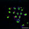
Kinda bored. Here's an overlay of three color fluorescence microscopy of human stem cells. By using a cocktail of growth factors, I can differentiate these cells into glutamatergic neurons...only takes about a week for them to completely change their shape. Action potential firing is observable after about 2 weeks. The ER is labeled with antibodies for Sec61b and a chimeric Sec61b protein that I designed. The nucleus shows fluorescence from nuclear localized mNeonGreen. Scale bar is 50 micrometers.

Here's another image of the endoplasmic reticulum in stem cells. This time I used antibodies against another protein I made and the protein called Vesicle Associated Membrane Protein Associated Protein B, VAPB

Here's an image of mitochondria in stem cells labeled with antibodies against my chimeric construct and the outer mitochondrial membrane protein TOMM20.


Here's another image of the endoplasmic reticulum in stem cells. This time I used antibodies against another protein I made and the protein called Vesicle Associated Membrane Protein Associated Protein B, VAPB

Here's an image of mitochondria in stem cells labeled with antibodies against my chimeric construct and the outer mitochondrial membrane protein TOMM20.

Last edited:


